A Comprehensive Review of Current and Emerging Approaches to Application of AI to Cardiology

Authors: Vivienne Hopkins, Yura Lee, Francis Lin, Hiba Rahman, & Vivash Rajan
Mentor: May Sin Ke. May is currently a doctoral candidate in the Department of Oncology at the University of Oxford.
Abstract
This review paper examines how the use of AI technology is gaining more significance in the field of cardiology, motivated by the reality that cardiovascular disease (CVD) continues to be the world’s leading cause of mortality, responsible for around 20 million deaths each year and costing an immense economic burden, totaling €282 billion yearly within the European Union. We discuss the applications of AI in cardiology medicine across the following areas. In diagnosis, AI models demonstrated strong performance across several major cardiovascular conditions, though their reliability varied depending on the disease and the metric used. A randomized trial found that AI guided ejection fraction assessment outperformed experienced sonographers, requiring fewer corrective revisions. In risk prediction, machine learning models outperformed traditional statistical approaches in classifying stroke and myocardial infarction risk. In terms of drug development, AI has played an important role in developing new compounds, performing cardio-toxicity screening, and repurposing existing drugs for use in cardiovascular applications. We highlighted several possible directions this research could go in the future, including clinical trials and validation studies that can help provide more clarity regarding how to take this research forward. Persistent limitations remain central to this discussion: many AI models function as uninterpretable “black boxes,” training data are frequently skewed toward wealthier populations with limited racial and ethnic diversity, and real world implementation studies show more modest gains than retrospective accuracy metrics suggest. We conclude that AI in cardiology currently functions best as a supportive tool for clinicians rather than an autonomous diagnostic system.
Introduction
Cardiovascular disease (CVD) refers to a group of diseases associated with the circulatory system, and is the number one cause of death, killing approximately 20 million people every year fig 1 (Naeem et al., 2026). As shown in Figure 1, CVD has a greater global mortality burden than any other disease category. There is a critical need to reduce mortality in CVDs and detecting the condition in its early stages and with greater accuracy is necessary to improve treatment outcomes for patients. In the present era, CVD is diagnosed with electrocardiography, cardiac MRI and CT scans, which allow for cardiovascular irregularities to be detected (Siontis et al., 2021). Despite this, with the growing complexity of CVD, artificial intelligence (AI) is proving to be a valuable asset to support and augment traditional approaches to cardiovascular diagnosis.
CVDs also add burden to lifelong social, economic burden on families and healthcare systems worldwide. Chronic pathologies such as ischemic heart disease and advanced heart failure are known to result in severe long-term functional disability, significantly reducing the patient’s quality of life and leading to broad, recurrent hospitalizations (Taylor et al., 2017). The failure to diagnose or treat these conditions can lead to progressive damage to myocardial tissue, resulting in irreversible ventricular remodeling, fatal heart failure, and unforeseen arrhythmias.
CVD creates a financial burden on society. In the European Union alone, it is estimated that managing CVD costs as much as €282 billion per year, which includes loss of income due to early exit from work, costs related to providing care to patients, and direct costs from medical treatment (Luengo-Fernandez et al., 2023). In lower income countries, treating cardiovascular conditions can surpass 100 percent of the national GDP per capita which can harshly limit access to care and negatively impact patient outcomes (Shakya et al., 2025). These issues increase the need for technologies to detect CVDs.

Common Cardiovascular Diseases
In this review we focus on 8 major CVDs as seen in Figure 2. Arrhythmias is an irregular heart beat and occurs when electric signals, that tell a heart to beat, aren’t functioning properly (Mayo Clinic, 2023). Acute myocardial ischemia occurs when some parts of the heart are not receiving enough oxygen rich blood (Mansoor-Beig et al., 2025). Myocardial infarction (MI), commonly known as a heart attack, is a severe consequence of acute myocardial ischemia, characterized by apoptosis, or programmed cell death, and impaired heart muscle function (Cicek & Bagci, 2024). Left ventricular dysfunction or LV dysfunction is when there is an inefficient delivery of blood flow from the main chamber of the heart to vital organs (Chahine & Alvey, 2023). Coronary artery disease (CAD) occurs when there is a build up of plaque in the heart's arteries (American Heart Association, 2024). A stroke results from disrupted blood flow to the brain and can be caused by a blocked cerebral artery or a ruptured artery (Khalafi et al., 2025). Heart failure is when the heart is unable to provide sufficient blood flow throughout the whole body, causing fluid to build up in the lungs (Mayo Clinic, 2025). Finally, left ventricular systolic dysfunction or LVSD, is when the left ventricle is too weak to pump blood out to your body (Cleveland Clinic, 2021).

Limitations in current standard of care
Despite advances in cardiovascular medicine, limitations remain in the current standards of care. Many CVDs develop gradually, resulting in delayed diagnosis, missed opportunities for early intervention, increased financial burdens and inaccurate assessment of patient risks (McClellan et al., 2019). Additionally, most cardiovascular research uses data that focuses on certain populations, leading to low representation of women, and racial and ethnic minorities, causing many physicians to struggle with understanding the extent of CVDs and symptoms (Sia & Poh, 2024).
Certain cardiovascular conditions remain difficult to diagnose. For example, transthyretin amyloid cardiomyopathy (ATTR-CM), a disease that causes heart muscles to stiffen, is often misdiagnosed because its symptoms resemble those of more common cardiac diseases (Rozenbaum et al., 2021). As a result, treatment is often delayed for patients. Additionally there is a lack of new disease treatments for CVDs with only one new drug developed in 2017 (McClellan et al., 2019).
Current cardiac medicine relies heavily on testing and imaging like ECGs and blood tests. However, some machines like ECGs can miss small details such as early-stage conduction defects that carry significant clinical risk if left untreated (Patel et al., 2025a). Also not all hospitals and clinics can house all of these tools. Most need special training and special equipment that hospitals with less funding have trouble acquiring (Patel et al., 2025b). Also, before giving back test results doctors often need to look at multiple outcomes of testing before reaching a diagnosis. This can lead to delayed diagnosis and missed opportunity for early solutions caused by human error, since analyzing the data takes time and is complicated (Patel et al., 2025b).
These limitations highlight several research gaps including the need for earlier and more accurate diagnosis, improved prediction of CVD risks, inclusive clinical data, and cost effective strategies - in which AI could potentially help improve. The scope of this review is to address these gaps and provide a strong rationale for the use of AI as a tool to enhance diagnosis, risk stratification, and personalized patient care while also diving into its limitations.
What is Artificial Intelligence
Artificial intelligence (AI) is the ability of a computer or a robot to perform intellectual tasks (McCarthy, 2007). The field of AI includes a variety of techniques, one of which is machine learning (ML). Scientists define machine learning as a set of methods that allow a computer to learn on its own without being programmed explicitly. There are two of the principal paradigms of ML: supervised and unsupervised. Within the framework of supervised learning, researchers use a set of labeled data for training algorithms so that the computer can establish patterns and make predictions (Topol, 2019). In turn, unsupervised learning works with an unlabeled data set to find patterns and connections. The use of these methods in health care is important because they allow analyzing large amounts of information and contribute to the process of making medical decisions (Topol, 2019).
A more sophisticated branch of machine learning is known as deep learning (DL). These networks are composed of multiple layers that enable DL to process big sets of data and detect patterns that may not be identified by traditional algorithms (LeCun et al., 2015). The use of DL in medical image analysis is appropriate because it can recognize patterns in images such as MRI and CT scans. For instance, in cardiology, DL can be utilized to make sense of cardiovascular images and signals to help diagnose disease and facilitate treatment (Litjens et al., 2017).
In order to enhance long-term survival rates and eliminate the negative impact of the aforementioned conditions, it is necessary to adopt new diagnostic technologies of fast and non-invasive character. Previously, the evaluation of heart structure was done through various imaging and electrophysiological instruments, which allowed choosing proper therapy methods.
An electrocardiogram (ECG) is a harmless procedure that records the electrical activity of the heart with electrodes (pads) attached to the skin (Kligfield et al., 2007). It is employed as a tool for diagnosis of arrhythmia, myocardial infarction, coronary artery disease and other disorders. A recent study found that AI could identify minute deviations from normal heart activity present in an ECG more accurately than traditional methods of diagnosis (Attia et al., 2019). Cardiac MRI is employed to obtain detailed information about the cardiac anatomy and rhythm, myocardial infarction, myocardial fibrosis and other abnormal conditions. However, it is costly and time consuming. AI simplifies cardiac MRI, which leads to faster scans and better image quality to enhance diagnostic accuracy (Oksuz et al., 2019). In the same way, CT scans are taken to obtain detailed images of the heart and to identify coronary plaque, heart aneurysm and other abnormal conditions of the cardiovascular system. However, they involve patients being exposed to harmful X-ray radiation (Einstein, 2012). AI in cardiac CT scans enhances diagnostic precision and aids in cardiovascular risk assessment, such as coronary plaque volume (Commandeur et al., 2018).
AI is also contributing to the improvement of cardiovascular diagnostics accuracy and to the ability to predict the risk of cardiovascular death. Specifically, Cho et al. (2024) developed an AI system with a very high predictive accuracy of cardiac death based on ECG data. It predicted, based on age and sex, other medical conditions, and the extent of heart failure, if the patient had any. Moreover, a recent study discovered that cardiologists using AI detected more cardiovascular irregularities, including those in high-risk patients, than those not using the technology (Tsai et al., 2025). These findings collectively illustrate how AI could potentially enhance cardiovascular diagnosis, benefiting doctors in clinical workplaces, and improving risk prediction, therefore making it a powerful tool in cardiology.
Current Applications of AI in Cardiology
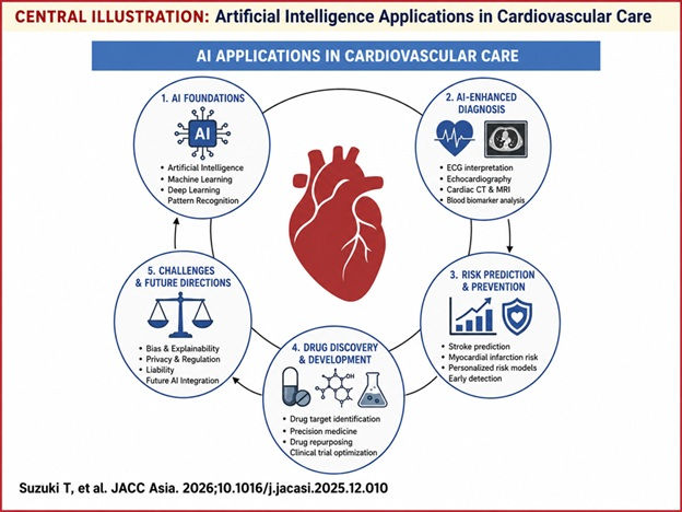
AI has reached every stage of the cardiology workflow, though unevenly. Natural language processing can pull risk factors and diagnoses out of free-text clinical notes (Reading-Turchioe et al., 2022). AI-enabled stethoscopes read single-lead ECG and heart sounds during routine auscultation and detect reduced ejection fraction in fifteen seconds (Bachtiger et al., 2022). ECG is furthest along. CNNs trained on 12-lead recordings detect left ventricular systolic dysfunction (LVSD) with an area under curve (AUC) of 0.93 (Attia et al., 2019), estimate left ventricular filling pressures (Lee et al., 2024), and identify patterns invisible to human readers (Siontis et al., 2021). The same recordings carry prognostic information: an AI-derived ECF score predicted in-hospital cardiac death in acute heart failure independently of age, sex, comorbidities and ejection fraction (Cho et al., 2024). In echocardiography, AI measures LVEF more consistently than experienced sonographers (He et al., 2023). Deep learning also flags motion-corrupted cardiac MRI scans that would otherwise need manual review (Oksuz et al., 2019), and quantifies epicardial fat from routine calcium-scoring CT for risk stratification (Commandeur et al., 2018). Biomarker interpretation is shifting from fixed thresholds toward individual estimates: CoDE-ACS combines troponin with clinical variables to give each patient a probability of myocardial infarction, and it outperforms the thresholds in current guidelines (Doudesis et al., 2023).
Results
In this section we will dive into how AI can help with cardiovascular care and prevention. AI has made huge strides in diagnosing complex CVDs, analyzing risk prediction models, and aiding in drug developments that can help cardiovascular medicine. We will also dive into the challenges, limitations and consequences of the use of AI in cardiology.
Diagnosis
One thing that researchers are studying with AI is its ability to help diagnosis. From acute myocardial ischemia to left ventricular dysfunction AI has shown promise in aiding doctors when diagnosing patients (see Table 1 below).
In a research study that included 30 studies looking at acute myocardial ischemia, which can lead to heart failure, in one of the reported studies they found that the AI was able to achieve a 92% specificity in a study with 30 different studies (Mansoor-Beig et al., 2025). In a study with 7,313 patients the AI achieved a 98.9% specificity and in a study with 8,493 patients the AI achieved a 84.8% specificity (Al-Zaiti et al., 2023; Lee et al., 2025). Specificity refers to the percent of people that the AI was able to correctly say that there was no acute myocardial ischemia. From the three papers the AI was able to get over 80% in specificity consistently.
In another review paper researchers looked at 9 studies that included 2,263 patients and their topic focused on coronary artery disease (CAD) (B. Liu et al., 2025). The AI scored a 94% sensitivity which measures the percent of people that the Ai was able to correctly find CAD in (B. Liu et al., 2025). As well individually between the different studies the AIs success rate fluctuated. These differences may have come from different AI algorithms, different imaging methods, or different ways of applying the AI (B. Liu et al., 2025). In a study of 2,303 patients using 12,954 ECG scans, an AI model achieved a 0.87 AUC score on detecting CAD, AUC=1 means perfect accuracy and 0.5 indicates random guessing, (Huang et al., 2022). Between the high sensitivity score and the high AUC score the AIs in both studies performed well.
A randomized clinical trial with a total of 3,495 studies, analyzed the superiority of AI guided workflow over traditional sonographic workflow when analyzing LVEF measurements in cardiac function assessments. The AI powered workflow significantly outperformed the traditional sonographic workflow with 16.8% of AI assisted studies needing large revisions while 27.2% of sonographic assisted studies needed large revisions (He et al., 2023). This trial was similarly done with the detection of left ventricular diastolic function and LVEF in 274,710 patients (Lee et al., 2024). The AI demonstrated excellent diagnostic accuracy and achieved a 95% confidence interval (Lee et al., 2024). It also correctly identified approximately 83% percent of patients with elevated filling pressures (Lee et al., 2024).
Lastly, a review group looked at 7 different research groups and found that between the 7 AIs that those researchers used attained a 83.3% sensitivity score and a 87% specificity score (Bjerkén et al., 2022). Another important factor is that the AI worked even between different ages, sexes, and other medical problems already identified in the patients (Bjerkén et al., 2022). In another study conducted of 1,996 adults 45 or older only 22 actually had left ventricular dysfunction and the AI spotted 19 of those 22 and correctly identified that 1,825 of the patients did not have left ventricular dysfunction additionally a study that collected 42,291 ECGs from 31,944 the AI has an AUC of 0.971 (Kashou et al., 2021; König et al., 2023). Across these studies, different AI models showed varying sensitivity and specificity for LV dysfunction. Even so the AIs as a whole scored above 80% throughout all of them and were close to hitting perfect accuracy in one of them.
Diagnostic performance of ECG, clinical data, and CT imaging for acute myocardial ischemia, coronary artery disease, and left ventricular dysfunction. AUC, area under the curve, evaluates the performance of AI and gives it a score, 1.0 perfect prediction accuracy, 0.5 random guessing, < 0.5 worse than random guessing; CAD, coronary artery disease, ECG, electrocardiogram, CT, computed tomography.
Table 1. A comparison of AI models between different CVDs.
AI Diagnosis Application | Acute Myocardial Ischemia | Coronary Artery Disease (CAD) | Left Ventricular Dysfunction |
ECG | (Herman et al., 2023) AUC of 0.938 AI can identify patterns in patients ECG scans that could show heart failure. | (Huang et al., 2022) AUC of 0.869 AI can analyze patterns in the electrical signals from the ECG scans that are realtor to CAD. | (König et al., 2023) AUC of 0.971 Like heart failure and CAD, AI can search for patterns in ECGs that could reveal an earlier stage of (LV) dysfunction or a later state of it. |
Clinical Data | (W.-C. Liu et al., 2021a) AUC of 0.978 AI can look through symptoms, past medical history, test results, and other information gathered to find patterns or other indications of heart failure or future heart failure/ problems. | (Forrest et al., 2022) AUC of 0.91 AI can combine potential risk factors and patient information to make an estimate of the chance of CAD. | (Y.-C. Huang et al., 2023) AUC of 0.85 AI can use information from the patients like age, previous heart disease, blood test results, and other findings to estimate the patients chance of having or in the future having Left ventricular dysfunction. |
CT Imaging | (Karlsberg et al., 2024) AUC of 0.85 AI can analyze CT imaging looking for problems with heart structures and heart functions that could be associated with heart failure. | (Bernardo et al., 2025) AUC of 0.91 per patient AI is able to analyze CT imaging by looking at the coronary artery and measure the narrowing of it. | (Bernardo et al., 2025) AUC of 0.96 AI can look over the CT Imaging and from the different sizes and shapes of different heart structures, and how much blood the heart is pumping to help make an educated guess. |
Risk Prediction
Risk prediction models enable earlier intervention, improve resource allocations and reduce healthcare costs (Cai et al., 2024). Recent advances in AI have enhanced these capabilities by integrating large, complex, and multi-model datasets to uncover patterns that may not be apparent through traditional approaches. Consequently, AI risk prediction models have emerged as a promising strategy for improving diagnostic accuracy and individualized cardiovascular care.
In reference to strokes, supervised machine learning models can rapidly analyze complex clinical datasets, reducing the time required for risk assessments while improving predictive accuracy (Heseltine-Carp et al., 2025). A study done with 4 different machine learning algorithms, naive Bayes, J48, K-nearest neighbor and random forest, showcased how new and advanced AI machine learning models outperformed old ones by speed and exhibited an accuracy of 99.8% in J48, K-nearest neighbor and random forest algorithms over 85.6% in the naive Bayes model (Dritsas & Trigka, 2022). AI has also expanded into wearable devices, including, ultrasonic neckbraces, ECG devices and even apps such as Stroke Riskometer as seen in Figure 4 (Chen & Sawan, 2021). Additional wearable devices such as smartwatches, fitness trackers and smart clothing use photoplethysmography (PPG) for physiological signals (Dagher et al., 2020). PPG sensors can monitor blood volume changes in any part of the body and measure subtle or alarming changes to the user or the clinician (Dagher et al., 2020). In addition, AI-assisted retinal imaging was created. When a patient's retinal morphology changes, like the thinning of the retinal nerve fiber or retinal ganglion cell layer, the AI will take that image and provide a risk stratification, or risk report, on how that change may lead to a stroke (Khalafi et al., 2025). These noninvasive, portable and cost-effective technologies have potential for improving stroke detection and prevention. (Khalafi et al., 2025).

AI has also demonstrated substantial value in predicting MI. In a clinical trial in the UK an AI logistical regression model and XGBoost machines were evaluated for MI risk prediction. This trial was done on over 500,000 patients in the course of approximately 13 years (Moore & Bell, 2022). Logistic regression models are created to monitor risk factors and are used when the outcome has two possible categories such as high risk vs low risk of CVD (Moore & Bell, 2022). The model combines multiple patient predictors, age, BMI, blood type, etc, to estimate the probability that a patient belongs to one category (Moore & Bell, 2022). XGBoost or extreme gradient boost, combines multiple decision trees, or tools for classifying diseases, to identify complex patterns in data, using each new tree to correct old data and evolve to a highly accurate model (Moore & Bell, 2022). Although both approaches improved early detection of individuals at risk for MI, XGBoost demonstrated superior predictive performance as seen below in Table 2 (Moore & Bell, 2022):

Similarly, a test done in Tri-General Hospital in Taipei, Taiwan, used 25,002 patients and a control group of 14,296 patients to test 12 ECGs that detected MI (W.-C Liu et al., 2021a). In the trial the AI model detected MI with a precision and sensitivity of 93.2%, showcasing both a fast and reliable AI risk prediction model (W.-C Liu et al., 2021b). Overall, AI-based cardiovascular risk prediction models improve the early detection of stroke and MI by enabling more accurate, personalized and timely assessments. These advances support earlier clinical interventions, optimize healthcare resources, and have the potential to reduce CVD mortality rates.
Drug Development
Most AI in cardiology works on diagnosis. Drug development is an aspect harder to judge, where trials run for years, most candidates fail, and software usually acts long before any result reaches a patient. Three uses have reached patients or approved drugs—designing new molecules, screening drugs for harm to the heart, and repurposing existing ones (Abdelhamid et al., 2026).
One trial tested whether AI could design a drug outright. Rentosertib was given to 71 patients with idiopathic pulmonary fibrosis, a scarring lung disease, at 22 sites in China (Xu et al., 2025). AI chose the biological target and designed the molecule. Patients on the highest dose showed improved lung function after 12 weeks while those on placebo declined, though the trial was small and short (Xu et al., 2025). It is a lung drug rather than a heart one, but the authors describe it as the first with both an AI-chosen target and an AI-designed structure to reach patients and report a benefit.
A second use targets the heart directly. Certain drugs block hERG, a channel that helps regulate heart rhythm, and can set off dangerous irregular heartbeats, a frequent reason drugs are withdrawn. A model called deephERG, trained on nearly 8,000 compounds, achieved a validation AUC of 0.967, outperforming a single-task neural network, a support vector machine, and a naive Bayesian classifier tested on the same data (Cai et al., 2019). Running across 1,824 approved drugs, it flagged 539 (29.6%) as possible hERG blockers; among 49 anticancer and immunomodulating drugs examined separately, 15 predicted blockers had supporting clinical or preclinical evidence of hERG-related effects (Cai et al., 2019). A later ensemble model, CardioTox net, achieved accuracies of 81%, 75.5% and 74.6% across three external test sets and generally outperformed DeepHIT, CardPred, two OCHEM predictors, and Pred-hERG 4.2 across several evaluation measures (Karim et al., 2021). This kind of check runs before a drug reaches a person, though it predicts a laboratory measure rather than an actual arrhythmia.
A third use is repurposing, or finding new disease for drugs that already exist. One system emulated clinical trials inside the health records of 1.2 million patients with coronary artery disease and flagged six drugs linked to better outcomes but not prescribed for it, all of them already cardiovascular medicines (R. Liu et al., 2021). A similar approach appeared during COVID-19, when a company’s AI screened existing drugs and pointed to baricitinib, an arthritis pill (Richardson et al., 2020). Randomized trials afterward reported shorter recovery and, in one, fewer deaths, and regulators authorized it (Kalil et al., 2021; Marconi et al., 2021). Here, AI identified a possible new use for an existing drug, which still had to be tested in randomized clinical trials.
Lastly, AI is also being used in testing drugs. Clinical trials are slow and costly, and much of that cost goes into recruiting a control group. AI offers a partial shortcut called a synthetic control arm, where a model trained on past patient records stands in for some of the real control patients, letting a trial enrol fewer of them (Thangaraj et al., 2024). Related models can take the outcome of one trial and estimate what it would have been in a different set of patients; in one case a model bridged two large blood-pressure trials, SPRINT and ACCORD, reproducing the effect seen in each from the other’s data (Thangaraj et al., 2024). For now these methods are early. The synthetic-control studies are proof-of-concept, reproducing the survival patterns of real control groups but not always matching the makeup of the real patients.

Challenges and limitations of AI
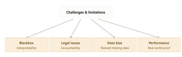
Though AI has been treated as a promising technology in cardiovascular medicine, there are many challenges at present to its clinical application (Figure 6). The technology has a number of theoretical advantages for diagnosis, prognosis and management of heart disease, but there are a number of significant issues to deal with – including lack of interpretability, potential for bias, liability and privacy/regulatory concerns – before it can be widely adopted in health care. As these difficulties come to light, the critical importance of clear algorithms, comprehensive regulatory protocols, and ongoing oversight by physicians becomes increasingly evident, underscoring the need for the safe and ethical application of AI in cardiovascular care.
The first constraint of AI for cardiovascular medicine is that it is not interpretable. In many instances, the actual decision making process of AI algorithms is a “black box” with the question of “how” or “why” a particular algorithm chooses one option over another remains unanswered. For example, many forms of AI will easily be able to discern patterns in data such as ECG, medical images, etc., which aren't quite as easy for humans (Patel et al., 2025a). But it's not always clear how an algorithm reached a certain conclusion, nor is it necessarily clear whether or not the algorithm's answer is correct. If these technologies were used in medical practice, it could result in serious misdiagnosis, and poor patient outcomes. There are evidently some researchers who believe that visualization tools, like Grad-CAM for an ECG or a medical image, can show users the portion of the image used for a specific conclusion, which can help users understand the rationale behind the AI’s output, but often lacks the information needed (Raghunath et al., 2020). It may become difficult for doctors to comprehend the reasoning behind the recommendation and they may not be able to adhere to all of the AI's recommendations.
The second challenge revolves around the legal liability issues surrounding the application of AI in cardiovascular medicine. Although most researchers agree that AI must be used as an assistive device and that there is no point in replacing doctors, there are still possibilities for diagnostic error, which can have detrimental impacts on patients. When the AI recommendation causes an incorrect diagnosis, it's hard to determine if this is a fault of the physician, the hospital, the software or the AI manufacturer. This issue is further complicated by the fact that in some instances, doctors might have diverging opinions with AI recommendations or might opt to not act on the recommendations at all (Patel et al., 2025b). Meanwhile, doctors would also be liable if they don't take an AI recommendation into account that they could have helped a patient with. Therefore, ethical and legal issues need to be seriously addressed when applying these technologies to medicine.
Next, data bias is a problem. Many cardiovascular AI systems are trained on data from white, affluent, and high income populations so they may perform less accurately for racial minorities, lower income patients, older adults, and people in low- and middle- income countries (Attia et al., 2019; Chamsi-Pasha & Chamsi-Pasha, 2025; Siontis et al., 2021). Their performance can also decline when applied in hospitals or populations different from those used for training (Poterucha et al., 2025). Unequal access to technologies such as smartwatches further limits representative data collection and may widen existing disparities (Chamsi-Pasha & Chamsi-Pasha, 2025). Unless these problems are addressed through more diverse datasets and external validation, cardiovascular AI would worsen rather than reduce health inequities.
Last but not least, its performance in the clinic is also a major problem. In particular, there are a number of algorithms which are performing poorly in predictions with regular patients. Many algorithms perform very well with a common measure of performance like the area under the receiver operating characteristic curve but have only a modest performance when measured on their overall accuracy. For instance, while an AI tool for detecting LV dysfunction had strong overall performance, the positive predictive value was only 33.8%, which means that more than one-third of patients who tested positive for the condition did not actually have it (Attia et al., 2019). In these situations patients' would have to suffer from unnecessary tests. Yet another problem is that the majority of AI algorithms provide a limited performance of detecting uncommon CVDs, since there are fewer examples for these diseases to be trained on (Gala et al., 2024). Cardiovascular AI applications, therefore, may not prove to be particularly useful in the real world because of their suboptimal performance features. For such measures, it is crucial that they are consistent for various populations, and that the algorithms generalize well to other settings.
Discussion
This review highlights several promising areas of research towards improving the early detection and management of CVDs. In summary, the studies reviewed here demonstrate that AI has evolved from a research curiosity to a measurable presence in virtually every diagnostic modality in cardiology. In imaging and electrocardiography, multiple models have equaled or outperformed humans under controlled circumstances: AI based LVEF assessment was superior to experienced sonographers (He et al., 2023). Part of what makes this finding compelling is its design, a blinded, randomized trial with nearly 3,500 echocardiograms, rather than the retrospective analyses that dominate most of this literature. A CNN trained on raw ECGs could detect severe LVSD with an AUC of 0.93 (Attia et al., 2019). These results support the central thesis of this review that AI can identify clinically important patterns in cardiac data that are missed during standard interpretation.
At the same time, the evidence has some unevenness that matters for how these findings should be understood. Diagnostic performance changes a lot depending on the disease and the metric used: AI models reached over 90% sensitivity for acute myocardial ischemia and coronary artery disease, but specificity for CAD dropped to 69% (W.-C Liu et al., 2025a), and even strong LVSD models had a positive predictive value low enough that most flagged patients would still need follow-up testing to confirm the diagnosis. Some of this variation likely reflects differences in study design rather than genuine differences in AI capability, the CAD meta analysis pooled heterogeneous imaging modalities and patient populations across multiple studies, while the LVSD model was validated on ECG echocardiogram pairs from a single large health system, conditions that tend to produce higher apparent accuracy than more diverse, multi-site settings would. This gap between high accuracy numbers and real-world reliability shows up in almost every section of this review, and it is the clearest common thread connecting diagnosis, risk prediction, and drug repurposing.
The real-world implementation data reinforce this caution. When AI ECG was used within a real hospital workflow instead of being tested after the fact, the absolute improvement over standard care was modest. The detection rates for unrecognized low ejection fraction were 1.5% compared to 1.1% (Tsai et al., 2025). This shows a meaningful improvement, but it is much smaller than what AUC based comparisons alone would indicate. Drug development illustrates a similar trend. There have been promising early results, but no AI designed cardiovascular drug has completed late stage testing (Xu et al., 2025).
In every application area, the pattern holds steady: AI excels as a tool for pattern recognition and support, but the research does not yet show it can operate without clinician oversight. The strongest studies here are the ones that treat AI as a second reader rather than a replacement, and that framing, supportive rather than autonomous, is likely to remain the operative model until prospective outcome trials, broader population validation, and clearer regulatory pathways close the gap between diagnostic accuracy and demonstrated clinical benefit.
Future research
The clearest priority for the field is to show that acting on an AI prediction improves how patients actually do. Most evidence gathered so far measures accuracy, meaning how often a model agrees with a scan or a later diagnosis, rather than whether using it lowers mortality or spares a patient an unnecessary test. Closing that gap will take prospective, pragmatic randomized trials that hand clinicians the tool and then track real outcomes over time (Bachtiger et al., 2022; Siontis et al., 2021). A few such trials have begun, but they remain the exception. Until more are done, more AI-cardiology tools will stay closer to research than to routine care.
A second priority is making models work outside the population that built them. Many AI ECG systems were trained on cohorts that are overwhelmingly white and drawn from wealthy health systems, and their accuracy tends to slip when applied elsewhere (Siontis et al., 2021). The fix depends less on smarter algorithms than on better data: cohorts that include women, older adults, and patients from the low- and middle- income countries where most CVDs actually occur. Federated learning, which trains a shared model across hospitals without moving patient records between them, offers one way to reach that breadth while keeping the data where it belongs (Thangaraj et al., 2024).
Integration is the next step. Today most models read a single signal, an ECG or a set of lab values, in isolation. The heart does not present itself one channel at a time, and the more promising direction fuses these streams with wearable and genomic data into models that see a patient whole. Digital twins are the ambitious end of this idea, which is a running simulation of an individual’s heart that updates as new data arrive and forecasts how it will respond to a given treatment (Thangaraj et al., 2024). Whether that vision is reachable will depend on unglamorous infrastructure, from standardized data formats to the wiring that connects an algorithm to a medical record, as much as on the models.
Drug development will be watched for a different milestone. The first AI designed drug has moved into late stage testing, yet none aimed at a cardiovascular condition has completed it. The coming years should show whether the early speed of AI discovery survives the harder test of late phase efficacy, and whether a heart drug is among the survivors. Underneath all of this sits governance. Rules on liability, privacy, and approval are still being written, and they will shape which tools reach patients as much as any technical advance. For all the progress in teaching machines to read a heart, the future lies in improving input data quality and building the evidence and gaps in knowledge that the AI model would depend on.
Conclusion
In this review, we provide a summary of AI and its effects on cardiology - from the current applications of AI in the field to its challenges and limitations. AI is used to help doctors diagnose patients by looking for patterns or problems with data or imaging. It is being used in many different forms of applications from echocardiography to AI enables stethoscopes to help find different common cardiovascular diseases like heart failure, stroke, LVSD, arrhythmias and many others. It has also been tested under risk prediction to see if it can aid doctors when they are working with patients. It has been studied to help catch problems before they get worse or by finding the risk of patients. AI has been studied to see if it can test drugs, make drugs, and find other ways a current drug can help other diseases. Even so, there are still limitations from the AI that will need to be studied and researched before AI can be implemented into the clinic.
References
Abdelhamid, A., Nasrallah, D., Al-Haneedi, Y., Ahmed, E., & Eid, A. H. (2026). The Emerging Role of Artificial Intelligence in Drug Discovery and Development: Implications for Cardiovascular Pharmacology. Journal of Cardiovascular Pharmacology, 87(6), 368–386. https://doi.org/10.1097/FJC.0000000000001803
Al-Zaiti, S. S., Martin-Gill, C., Zègre-Hemsey, J. K., Bouzid, Z., Faramand, Z., Alrawashdeh, M. O., Gregg, R. E., Helman, S., Riek, N. T., Kraevsky-Phillips, K., Clermont, G., Akcakaya, M., Sereika, S. M., Van Dam, P., Smith, S. W., Birnbaum, Y., Saba, S., Sejdic, E., & Callaway, C. W. (2023). Machine learning for ECG diagnosis and risk stratification of occlusion myocardial infarction. Nature Medicine, 29, 1–10. https://doi.org/10.1038/s41591-023-02396-3
American Heart Association. (2024). Coronary Artery Disease - coronary Heart Disease. https://www.heart.org/en/health-topics/consumer-healthcare/what-is-cardiovascular-disease/coronary-artery-disease
Attia, Z. I., Kapa, S., Lopez-Jimenez, F., McKie, P. M., Ladewig, D. J., Satam, G., Pellikka, P. A., Enriquez-Sarano, M., Noseworthy, P. A., Munger, T. M., Asirvatham, S. J., Scott, C. G., Carter, R. E., & Friedman, P. A. (2019). Screening for cardiac contractile dysfunction using an artificial intelligence-enabled electrocardiogram. Nature Medicine, 25(1), 70–74. https://doi.org/10.1038/s41591-018-0240-2
Bachtiger, P., Petri, C. F., Scott, F. E., Park, S. R., Kelshiker, M. A., Sahemey, H. K., Dretzke, J., Pabari, P. A., Halcox, J. P. J., Nicol, E. D., Francis, D. P., & Peters, N. S. (2022). Point-of-care screening for heart failure with reduced ejection fraction using artificial intelligence during ECG-enabled stethoscope examination in London, UK: A prospective, observational, multicentre study. The Lancet Digital Health, 4(2), e117–e125. https://doi.org/10.1016/S2589-7500(21)00256-9
Bernardo, R., Nurmohamed, N. S., Bom, M. J., Jukema, R., de Winter, R. W., Sprengers, R., Stroes, E. S. G., Min, J. K., Earls, J., Danad, I., Choi, A. D, & Knaapen, P. (2025). Diagnostic accuracy in coronary CT angiography analysis: Artificial intelligence versus human assessment. Open Heart, 12(1), e003115. https://doi.org/10.1136/openhrt-2024-003115
Bjerkén, L. V., Rønborg, S. N., Jensen, M. T., Ørting, S. N., & Nielsen, O. W. (2022). Artificial intelligence enabled ECG screening for left ventricular systolic dysfunction: A systematic review. Heart Failure Reviews, 28. https://doi.org/10.1007/s10741-022-10283-1
Cai, C., Guo, P., Zhou, Y., Zhou, J., Wang, Q., Zhang, F., Fang, J., & Cheng, F. (2019). Deep learning-based prediction of drug-induced cardiotoxicity. Journal of Chemical Information and Modeling, 59(3), 1073–1084. https://doi.org/10.1021/acs.jcim.8b00769
Cai, Y., Cai, Y.-Q., Tang, L.-Y., Wang, Y.-H., Gong, M., Jing, T.-C., Li, H.-J., Li-Ling, J., Hu, W., Yin, Z., Gong, Da X., & Zhang, G.-W. (2024). Artificial intelligence in the risk prediction models of cardiovascular disease and development of an independent validation screening tool: a systematic review. BMC Medicine, 22(1). https://doi.org/10.1186/s12916-024-03273-7
Chahine, J., & Alvey, H. (2023). Left Ventricular Failure. StatPearls Publishing. https://www.ncbi.nlm.nih.gov/books/NBK537098/
Chamsi-Pasha, M. A. R., & Chamsi-Pasha, H. (2025). Ethical challenges and current opportunities of artificial intelligence in cardiology. Journal of Cardiovascular Development and Disease, 15(3), 95–99. https://doi.org/10.1055/s-0045-1809879
Chen, Y.-H., & Sawan, M. (2021). Trends and Challenges of Wearable Multimodal Technologies for Stroke Risk Prediction. Sensors, 21(2), 460. https://doi.org/10.3390/s21020460
Cho, Y., Yoon, M., Kim, J., Lee, J. H., Oh, I.-Y., Lee, C. J., Kang, S.-M., & Choi, D.-J. (2024). Artificial intelligence–based electrocardiographic biomarker for outcome prediction in patients with acute heart failure. Journal of Medical Internet Research, 26, e52139. https://doi.org/10.2196/52139
Cicek, V., & Bagci, U. (2024). AI-powered contrast-free cardiovascular magnetic resonance imaging for myocardial infarction. Frontiers in Cardiovascular Medicine, 11. https://doi.org/10.3389/fcvm.2024.1457498
Cleveland Clinic. (2021). Left-sided heart failure: Symptoms, causes and treatment. https://my.clevelandclinic.org/health/diseases/22181-left-sided-heart-failure
Commandeur, F., Goeller, M., Betancur, J., Cadet, S., Doris, M., Chen, X., Berman, D. S., Slomka, P. J., Tamarappoo, B. K., & Dey, D. (2018). Deep learning for quantification of epicardial and thoracic adipose tissue from non-contrast CT. IEEE Transactions on Medical Imaging, 37(8), 1835–1846. https://doi.org/10.1109/TMI.2018.2804799
Dagher, L., Shi, H., Zhao, Y., & Marrouche, N. F. (2020). Wearables in cardiology: Here to stay. Heart Rhythm, 17(5), 889–895. https://doi.org/10.1016/j.hrthm.2020.02.023
Doudesis, D., Lee, K.K., Boeddinghaus, J. et al. (2023). Machine learning for diagnosis of myocardial infarction using cardiac troponin concentrations. Nature Medicine, 29(9), 1201–1210. https://doi.org/10.1038/s41591-023-02325-4
Dritsas, E., & Trigka, M. (2022). Stroke risk prediction with machine learning techniques. Sensors, 22(13), 4670. https://doi.org/10.3390/s22134670
Einstein, A. J. (2012). Effects of radiation exposure from cardiac imaging: How good are the data? Journal of the American College of Cardiology, 59(6), 553–565. https://doi.org/10.1016/j.jacc.2011.08.079
Forrest, I. S., Petrazzini, B. O., Duffy, Á., Park, J. K., Marquez-Luna, C., Jordan, D. M., Rocheleau, G., Cho, J. H., Rosenson, R. S., Narula, J., Nadkarni, G. N., & Do, R. (2022). Machine learning-based marker for coronary artery disease: Derivation and validation in two longitudinal cohorts. Lancet, 401(10372), 215. https://doi.org/10.1016/S0140-6736(22)02079-7
Gala, D., Behl, H., Shah, M., & Makaryus, A. N. (2024). The role of artificial intelligence in improving patient outcomes and future of healthcare delivery in cardiology: A narrative review of the literature. Healthcare, 12(4), 481. https://doi.org/10.3390/healthcare12040481
He, B., Kwan, A. C., Cho, J. H., Yuan, N., Pollick, C., Shiota, T., Ebinger, J., Bello, N. A., Wei, J., Josan, K., Duffy, G., Jujjavarapu, M., Siegel, R., Cheng, S., Zou, J. Y., & Ouyang, D. (2023). Blinded, randomized trial of sonographer versus AI cardiac function assessment. Nature, 616(7957), 520–524. https://doi.org/10.1038/s41586-023-05947-3
Hepzibah, K., & Priscila, S. S. (2024). Classification of Heart Diseases Using Logistic Regression with Various Preprocessing Techniques. Communications in Computer and Information Science, 83–95. https://doi.org/10.1007/978-3-031-59097-9_7
Herman, R., H Pendell Meyers, Smith, S. W., Bertolone, D. T., Leone, A., Konstantinos Bermpeis, Viscusi, M. M., Belmonte, M., Demolder, A., Boza, V., Vavrik, B., Viera Kresnakova, Andrej Iring, Martonak, M., Jakub Bahyl, Timea Kisova, Schelfaut, D., Vanderheyden, M., Perl, L., … Barbato, E. (2023). International evaluation of an artificial intelligence-powered ecg model detecting acute coronary occlusion myocardial infarction. European Heart Journal– Digital Health, 5(2). https://doi.org/10.1093/ehjdh/ztad074
Heseltine-Carp, W., Courtman, M., Browning, D., Kasabe, A., Allen, M., Streeter, A., Ifeachor, E., James, M., & Mullin, S. (2025). Machine learning to predict stroke risk from routine hospital data: A systematic review. International Journal of Medical Informatics, 196, 105811. https://doi.org/10.1016/j.ijmedinf.2025.105811
Huang, P.-S., Tseng, Y.-H., Tsai, C.-F., Chen, J.-J., Yang, S.-C., Chiu, F.-C., Chen, Z.-W., Hwang, J.-J., Chuang, E. Y., Wang, Y.-C., & Tsai, C.-T. (2022). An Artificial Intelligence-Enabled ECG Algorithm for the Prediction and Localization of Angiography-Proven Coronary Artery Disease. Biomedicines, 10(2), 394. https://doi.org/10.3390/biomedicines10020394
Huang, Y.-C., Hsu, Y.-C., Liu, Z.-Y., Lin, C.-H., Tsai, R., Chen, J.-S., Chang, P.-C., Liu, H.-T., Lee, W.-C., Wo, H.-T., Chou, C.-C., Wang, C.-C., Wen, M.-S., & Kuo, C.-F. (2023). Artificial intelligence-enabled electrocardiographic screening for left ventricular systolic dysfunction and mortality risk prediction. Frontiers in Cardiovascular Medicine, 10, 1070641. https://doi.org/10.3389/fcvm.2023.1070641
Kalil, A. C., Patterson, T. F., Mehta, A. K., Tomashek, K. M., Wolfe, C. R., Ghazaryan, V., Marconi, V. C., Ruiz-Palacios, G. M., Hsieh, L., Kline, S., Tapson, V., Iovine, N. M., Jain, M. K., Sweeney, D. A., El Sahly, H. M., Branche, A. R., Regalado Pineda, J., Lye, D. C., Sandkovsky, U., Luetkemeyer, A. F., … ACTT-2 Study Group Members (2021). Baricitinib plus Remdesivir for Hospitalized Adults with Covid-19. The New England Journal of Medicine, 384(9), 795–807. https://doi.org/10.1056/NEJMoa2031994
Karim, A., Lee, M., Balle, T., & Sattar, A. (2021). CardioTox net: a robust predictor for hERG channel blockade based on deep learning meta-feature ensembles. Journal of Cheminformatics, 13(1), 60. https://doi.org/10.1186/s13321-021-00541-z
Karlsberg P. R., S. Nurmohamed, N. S., Quesada, C. G., Samuels, B. A., Dohad, S., Anderson, L. R., Crabtree, T., Min, J. K., Choi, A. D., & Earls, J. P. (2024). Performance of an artificial intelligence-guided quantitative coronary computed tomography algorithm for predicting myocardial ischemia in real-world practice. IJC Heart & Vasculature, 53, 101433. https://doi.org/10.1016/j.ijcha.2024.101433
Kashou, A. H., Medina-Inojosa, J. R., Noseworthy, P. A., Rodeheffer, R. J., Lopez-Jimenez, F., Attia, I. Z., Kapa, S., Scott, C. G., Lee, A. T., Friedman, P. A., & McKie, P. M. (2021). Artificial Intelligence–Augmented Electrocardiogram Detection of Left Ventricular Systolic Dysfunction in the General Population. Mayo Clinic Proceedings, 96(10), 2576–2586. https://doi.org/10.1016/j.mayocp.2021.02.029
Khalafi, P., Morsali, S., Hamidi, S., Ashayeri, H., Sobhi, N., Pedrammehr, S., & Jafarizadeh, A. (2025). Artificial intelligence in stroke risk assessment and management via retinal imaging. Frontiers in Computational Neuroscience, 19, Article 1490603. https://doi.org/10.3389/fncom.2025.1490603
Kligfield, P., Gettes, L. S., Bailey, J. J., Childers, R., Deal, B. J., Hancock, E. W., van Herpen, G., Kors, J. A., Macfarlane, P., Mirvis, D. M., Pahlm, O., Rautaharju, P., & Wagner, G. S. (2007). Recommendations for the standardization and interpretation of the electrocardiogram: Part I: The electrocardiogram and its technology: A scientific statement from the American Heart Association Electrocardiography and Arrhythmias Committee, Council on Clinical Cardiology; the American College of Cardiology Foundation; and the Heart Rhythm Society. Circulation, 115(10), 1306–1324. https://doi.org/10.1161/CIRCULATIONAHA.106.180200
König, S., Hohenstein, S., Nitsche, A., Pellissier, V., Leiner, J., Stellmacher, L., Hindricks, G., & Bollmann, A. (2023). Artificial intelligence-based identification of left ventricular systolic dysfunction from 12-lead electrocardiograms: External validation and advanced application of an existing model. European Heart Journal - Digital Health, 5(2), 144–151. https://doi.org/10.1093/ehjdh/ztad081
Lee, E., Ito, S., Miranda, W. R., Lopez-Jimenez, F., Kane, G. C., Asirvatham, S. J., Noseworthy, P. A., Friedman, P. A., Carter, R. E., Borlaug, B. A., Attia, Z. I., & Oh, J. K. (2024). Artificial intelligence-enabled ECG for left ventricular diastolic function and filling pressure. Npj Digital Medicine, 7(4), 1–7. https://doi.org/10.1038/s41746-023-00993-7
Lee, M. S., Shin, T. G., Lee, Y., Kim, D. H., Choi, S. H., Cho, H., Lee, M. J., Jeong, K. Y., Kim, W. Y., Min, Y. G., Han, C., Yoon, J. C., Jung, E., Kim, W. J., Ahn, C., Seo, J. Y., Lim, T. H., Kim, J. S., Choi, J., … Jo, Y.-Y. (2025). Artificial intelligence applied to electrocardiogram to rule out acute myocardial infarction: The ROMIAE multicentre study. European Heart Journal, 46(20). https://doi.org/10.1093/eurheartj/ehaf004
LeCun, Y., Bengio, Y., & Hinton, G. (2015). Deep learning. Nature, 521(7553), 436–444. https://doi.org/https://doi.org/10.1038/nature14539
Litjens, G., Kooi, T., Bejnordi, B. E., Setio, A. A. A., Ciompi, F., Ghafoorian, M., van der Laak, J. A. W. M., van Ginneken, B., & Sánchez, C. I. (2017). A survey on deep learning in Medical image analysis. Medical Image Analysis, 42(1), 60–88. https://doi.org/10.1016/j.media.2017.07.005
Liu, B., Reis, J., Sharma, A., & Wang, W. (2025). Application of artificial intelligence in non-invasive cardiovascular imaging for coronary artery disease: A systematic review and meta-analysis [Review of Application of artificial intelligence in non-invasive cardiovascular imaging for coronary artery disease: A systematic review and meta-analysis]. Frontiers in Cardiovascular Medicine, 12. https://doi.org/10.3389/fcvm.2025.1664183
Liu, W.-C., Lin, C.-S., Tsai, C.-S., Tsao, T.-P., Cheng, C.-C., Liou, J.-T., Lin, W.-S., Cheng, S.-M., Lou, Y.-S., Lee, C.-C., & Lin, C. (2021a). A deep learning algorithm for detecting acute myocardial infarction. EuroIntervention, 17(9), 765–773. https://doi.org/10.4244/eij-d-20-01155
Liu, W.-C., Lin, C., Lin, C.-S., Tsai, M.-C., Chen, S.-J., Tsai, S.-H., Lin, W.-S., Lee, C.-C., Tsao, T.-P., & Cheng, C.-C. (2021b). An Artificial Intelligence-Based Alarm Strategy Facilitates Management of Acute Myocardial Infarction. Journal of Personalized Medicine, 11(11), 1149. https://doi.org/10.3390/jpm11111149
Liu, R., Wei, L., & Zhang, P. (2021). A deep learning framework for drug repurposing via emulating clinical trials on real-world patient data. Nature Machine Intelligence, 3(1), 68–75. https://doi.org/10.1038/s42256-020-00276-w
Luengo-Fernandez, R., Walli-Attaei, M., Gray, A., Torbica, A., Maggioni, A. P., Huculeci, R., Bairami, F., Aboyans, V., Timmis, A. D., Vardas, P., & Leal, J. (2023). Economic burden of cardiovascular diseases in the European Union: a population-based cost study. European Heart Journal, 44(45), 4752–4767. https://doi.org/10.1093/eurheartj/ehad583
Mansoor-Beig, S. T., Sheriff, M. M., Alhejaili, A. E. Z., Kamel, A. D. M., Elnair, S. I., Ahmed, M. A., Mawardi, L. M. H., Fida, L. A., Abdulhafeez, E., Jan, R. H., Tukruni, H. Y. I., Aljahdali, R. A., Alshahrani, K. Z. M., Hakami, W. H., & Saati, A. A. Z. (2025). The role of artificial intelligence in improving diagnosis, management, and outcomes of acute myocardial ischemia: A systematic review. Cureus, 17(12), e98865. https://doi.org/10.7759/cureus.98865
Marconi V., Ramanan A., de Bono S., Kartman, C. E., Krishnan, V., Liao, R., Piruzeli, M. L. B., Goldman, J. D., Alatorre-Alexander, J., de Cassia Pellegrini, R., Estrada, V., Som, M., Cardoso, A., Chakladar, S., Crowe, B., Reis, P., Zhang, X., Adams, D. H., Ely, E. W., & COV-BARRIER Study Group. (2021). Efficacy and safety of baricitinib for the treatment of hospitalised adults with COVID-19 (COV-BARRIER): a randomised, double-blind, parallel-group, placebo-controlled phase 3 trial. The Lancet Respiratory Medicine, 9, 1407-1418. https://doi.org/10.1016/S2213-2600(21)00331-3
Mayo Clinic. (2023). Heart arrhythmia. Mayo Clinic. https://www.mayoclinic.org/diseases-conditions/heart-arrhythmia/symptoms-causes/syc-20350668
McCarthy, J. (2007). What is artificial intelligence? Stanford University. http://jmc.stanford.edu/articles/whatisai/whatisai.pd
McClellan, M., Brown, N., Califf, R. M., & Warner, J. J. (2019). Call to Action: Urgent Challenges in Cardiovascular Disease: A Presidential Advisory From the American Heart Association. Circulation, 139(9). https://doi.org/10.1161/cir.0000000000000652
Moore, A., & Bell, M. (2022). XGBoost, A Novel Explainable AI Technique, in the Prediction of Myocardial Infarction: A UK Biobank Cohort Study. Clinical Medicine Insights: Cardiology, 16, 117954682211336. https://doi.org/10.1177/11795468221133611
Naeem, U., Deo, S., Sahebkar, A., Sheikh, S., Khoja, A., Vaughan, E. M., Abdul Jabbar, A. B., Kalra, D. K., Slipczuk, L., & Virani, S. S. (2026). Global burden of cardiovascular disease: What should be expected in the next 25 years? Current Cardiology Reports, 28(1), Article 31. https://doi.org/10.1007/s11886-026-02355-7
Oksuz, I., Ruijsink, B., Puyol-Antón, E., Clough, J. R., Cruz, G., Bustin, A., Prieto, C., Botnar, R., Rueckert, D., Schnabel, J. A., & King, A. P. (2019). Automatic CNN-based detection of cardiac MR motion artefacts using k-space data augmentation and curriculum learning. Medical Image Analysis, 55, 136–147. https://doi.org/10.1016/j.media.2019.04.009
Patel, D., Kantamneni, R., John, J. D., Patel, T., Shukla, A., Salma, A., & Anand, N. (2025a). Artificial intelligence in cardiology: an updated systematic review with ethical considerations and challenges in implementing artificial intelligence models. Annals of Medicine and Surgery, 88(2), 1789–1805. https://doi.org/10.1097/MS9.0000000000004607
Patel, D., Millis, R. M., Khan, S., Patel, T., Joshua, S., Khan, S., & Chetarajupalli, C. (2025b). A narrative review on ethical considerations and challenges in AI-driven cardiology. Annals of Medicine and Surgery, 87(7), 4152–4164. https://doi.org/10.1097/MS9.0000000000003349
Poterucha, T. J., Jing, L., Pimentel Ricart, R., Adjei-Mosi, M., Finer, J., Hartzel, D., Kelsey, C., Long, A., Rocha, D., Ruhl, J. A., vanMaanen, D., Probst, M. A., Daniels, B., Joshi, S. D., Tastet, O., Corbin, D., Avram, R., Barrios, J. P., Tison, G. H., ... Elias, P. (2025). Detecting structural heart disease from electrocardiograms using AI. Nature, 644(8078), 221–229. https://doi.org/10.1038/s41586-025-09227-0
Raghunath, S., Ulloa Cerna, A. E., Jing, L., vanMaanen, D. P., Stough, J., Hartzel, D. N., Leader, J. B., Kirchner, H. L., Stumpe, M. C., Hafez, A., Nemani, A., Carbonati, T., Johnson, K. W., Young, K., Good, C. W., Pfeifer, J. M., Patel, A. A., Delisle, B. P., Alsaid, A., … Fornwalt, B. K. (2020). Prediction of mortality from 12-lead electrocardiogram voltage data using a deep neural network. Nature Medicine, 26(6), 886–891. https://doi.org/10.1038/s41591-020-0870-z
Reading Turchioe, M., Volodarskiy, A., Pathak, J., Wright, D. N., Tcheng, J. E., & Slotwiner, D. (2022). Systematic review of current natural language processing methods and applications in cardiology. Heart (British Cardiac Society), 108(12), 909–916. https://doi.org/10.1136/heartjnl-2021-319769
Richardson, P., Griffin, I., Tucker, C., Smith, D., Oechsle, O., Phelan, A., Rawling, M., Savory, E., & Stebbing, J. (2020). Baricitinib as potential treatment for 2019-nCoV acute respiratory disease. Lancet, 395(10223), e30–e31. https://doi.org/10.1016/S0140-6736(20)30304-4
Rozenbaum, M. H., Large, S., Bhambri, R., Stewart, M., Whelan, J., van Doornewaard, A., Dasgupta, N., Masri, A., & Nativi-Nicolau, J. (2021). Impact of Delayed Diagnosis and Misdiagnosis for Patients with Transthyretin Amyloid Cardiomyopathy (ATTR-CM): A Targeted Literature Review. Springer Nature, 10(1), 141–159. https://doi.org/10.1007/s40119-021-00219-5
Shakya, S., Shrestha, A., Robinson, S., Randall, S., Mnatzaganian, G., Brown, H., Boyd, J., Xu, D., Lee, C. M. Y., Brumby, S., Peeters, A., Lucas, J., Gauci, S., Huxley, R., O'Neil, A., & Gao, L. (2025). Global comparison of the economic costs of coronary heart disease: a systematic review and meta-analysis. BMJ Open, 15(1), e084917. https://doi.org/10.1136/bmjopen-2024-084917
Sia, C.-H., & Poh, K.-K. (2024). Advances and challenges in cardiology. Singapore Medical Journal, 65(7), 369–369. https://doi.org/10.4103/singaporemedj.smj-2024-128
Siontis, K. C., Noseworthy, P. A., Attia, Z. I., & Friedman, P. A. (2021). Artificial intelligence-enhanced electrocardiography in cardiovascular disease management. Nature Reviews Cardiology, 18(7), 465–478. https://doi.org/10.1038/s41569-020-00503-2
Suzuki, T., & Suzuki, R. (2026). Artificial intelligence and digital innovation in cardiovascular medicine. JACC: Asia. Advance online publication. https://doi.org/10.1016/j.jacasi.2025.12.010
Taylor, C. J., Ryan, R., Nichols, L., Gale, N., Hobbs, F. D. R., & Marshall, T. (2017). Survival following a diagnosis of heart failure in primary care. Family Practice, 34(2), 161–168. https://doi.org/10.1093/fampra/cmw145
Thangaraj, P. M., Benson, S. H., Oikonomou, E. K., Asselbergs, F. W., & Khera, R. (2024). Cardiovascular care with digital twin technology in the era of generative artificial intelligence. European Heart Journal, 45(45), 4808–4821. https://doi.org/10.1093/eurheartj/ehae619
Topol, E. J. (2019). High-performance medicine: The convergence of human and artificial intelligence. Nature Medicine, 25(1), 44–56. https://doi.org/10.1038/s41591-018-0300-7
Tsai, D. J., Lin, C., Liu, W. T., Lee, C. C., Chang, C. H., Lin, W. Y., Liu, Y. L., Chang, D. W., Hsieh, P. H., Tsai, C. S., Chen, Y. H., Hung, Y. J., & Lin, C. S. (2025). Artificial intelligence-assisted diagnosis and prognostication in low ejection fraction using electrocardiograms in inpatient department: a pragmatic randomized controlled trial. BMC Medicine, 23(1), 342. https://doi.org/10.1186/s12916-025-04190-z
World Health Organization. (2025, July 31). Cardiovascular diseases (CVDs). https://www.who.int/news-room/fact-sheets/detail/cardiovascular-diseases-(cvds)
Xu, Z., Ren, F., Wang, P., Cao, J., Tan, C., Ma, D., Zhao, L., Dai, J., Ding, Y., Fang, H., Li, H., Liu, H., Luo, F., Meng, Y., Pan, P., Xiang, P., Xiao, Z., Rao, S., Satler, C., . . . Zhavoronkov, A. (2025). A generative AI-discovered TNIK inhibitor for idiopathic pulmonary fibrosis: a randomized phase 2a trial. Nature Medicine, 31, 2602–2610. doi:10.1038/s41591-025-03743-2


Comments